When I first began working with granulation tissue wound pictures in clinical care, I realized they told a story beyond what written notes could capture. These images don’t just document wounds—they highlight whether the body is actively rebuilding or signaling complications that need attention.
From my own experience, a wound showing moist, beefy-red tissue is often a reassuring sign of healthy blood vessel growth. In contrast, pale, friable, or uneven tissue can reveal poor circulation, stalled recovery, or even infection. Recognizing these distinctions early helps prevent setbacks and promotes better outcomes.
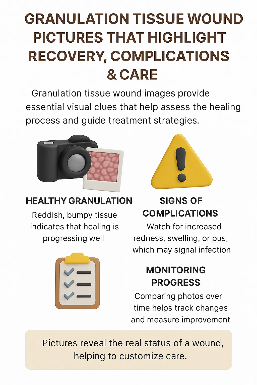
In this guide, you’ll learn how wound photos reveal signs of recovery, what complications to look for, and how these insights support better care for both patients and caregivers.
Top Takeaways
- Wound photos highlight recovery or setbacks by showing changes in tissue health.
- Red and moist tissue = good progress; pale or uneven = warning signs.
- Photo documentation improves outcomes by guiding care and motivating patients.
- Consistency builds trust—photos provide visible proof of healing.
- Action is key—use photos alongside medical guidance for safe recovery.
What Wound Pictures Reveal
Granulation tissue wound pictures provide visual evidence of healing.
- Signs of recovery: Healthy tissue looks moist, red or pink, and slightly bumpy, indicating good blood supply and collagen formation.
- Signs of complications: Pale, gray, dry, or irregular tissue may suggest infection, poor circulation, or delayed healing.
By comparing wound photos over time, patients and caregivers can see whether progress is steady or if care needs to be adjusted, making proper wound care an essential part of monitoring healing and ensuring the best possible outcomes.
Expert Insight
"In my wound care practice, photos are more than documentation—they are checkpoints. A healthy, moist red tissue bed signals active repair, while pale or uneven tissue warns of complications. Reading these images early can make the difference between smooth healing and prolonged recovery."
Case Studies & Real-World Examples
Post-Surgical Recovery
Patient: 54-year-old after abdominal surgery
Early signs: Pale, uneven tissue pointing to circulation issues
Interventions: Improved nutrition, better dressings, mobility support
Outcome: By week 4, wound photos revealed strong red granulation tissue
Takeaway: Visual progress reassured both the patient and care team
Diabetic Foot Ulcer
Patient: Chronic diabetic ulcer
Challenge: Slow recovery and friable tissue spotted in weekly photos
Interventions: Prompt debridement and infection control
Outcome: Photos showed the shift to healthier tissue within weeks
Takeaway: Photo tracking guided interventions and boosted patient confidence
Research & Clinical Perspective
Studies confirm wound photography supports better healing outcomes
From experience, photos act as teaching tools, helping patients recognize both healthy progress and warning signs
Patient: 54-year-old after abdominal surgery
Early signs: Pale, uneven tissue pointing to circulation issues
Interventions: Improved nutrition, better dressings, mobility support
Outcome: By week 4, wound photos revealed strong red granulation tissue
Takeaway: Visual progress reassured both the patient and care team
Patient: Chronic diabetic ulcer
Challenge: Slow recovery and friable tissue spotted in weekly photos
Interventions: Prompt debridement and infection control
Outcome: Photos showed the shift to healthier tissue within weeks
Takeaway: Photo tracking guided interventions and boosted patient confidence
Studies confirm wound photography supports better healing outcomes
From experience, photos act as teaching tools, helping patients recognize both healthy progress and warning signs
Supporting Statistics
Chronic wounds are costly and widespread
Over 8.2 million Medicare patients affected annually; costs range from $28–$96 billion
Source: NIH – Chronic Wounds Study
Pressure ulcers affect millions
More than 2.5 million Americans develop them each year
Source: AHRQ – Pressure Ulcer Facts
Diabetic foot ulcers are high-risk
12–25% of people with diabetes will develop a foot ulcer in their lifetime
Source: NCBI – Diabetic Foot Ulcers
Severe long-term impact
Recurrence: up to 65% within 5 years; amputation and mortality rates remain high
Source: American Diabetes Association – Foot Complications
Photography is clinically supported
Recognized as a valuable complement to written wound documentation
Source: PubMed – Wound Photography Guidelines
Final Thought & Opinion
Granulation tissue wound pictures are more than images—they highlight recovery milestones, potential complications, and care needs. They offer clarity when written descriptions fall short, giving patients motivation and providers actionable insights.
In my view, photos transform care by making healing visible. They inspire trust, encourage patients, and help providers act before complications escalate. Simply put, granulation tissue wound pictures are essential tools for recovery, complication prevention, and patient-centered care.
Next Steps
Check your wound photos: Healthy = red, moist tissue; warning = pale or uneven tissue
Track consistently: Same angle, lighting, and timing for accuracy
Share with professionals: Use photos as part of your care discussions
Use trusted resources: NIH, CDC, ADA
Act fast if needed: Seek professional help if photos reveal infection or slow recovery


